Cheek Cell Diagram Human Cheek Cell Dna Extraction
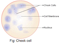
Draw the human cheek cell with correct labelling Cheek cell diagram Cheek cells under microscope labeled
Cheek Cells Under Microscope Labeled
Cheek cell lab – hailey's blog Cheek cell lab – filip’s blog Human cheek cell dna extraction
[diagram] human cheek cell diagram labeled
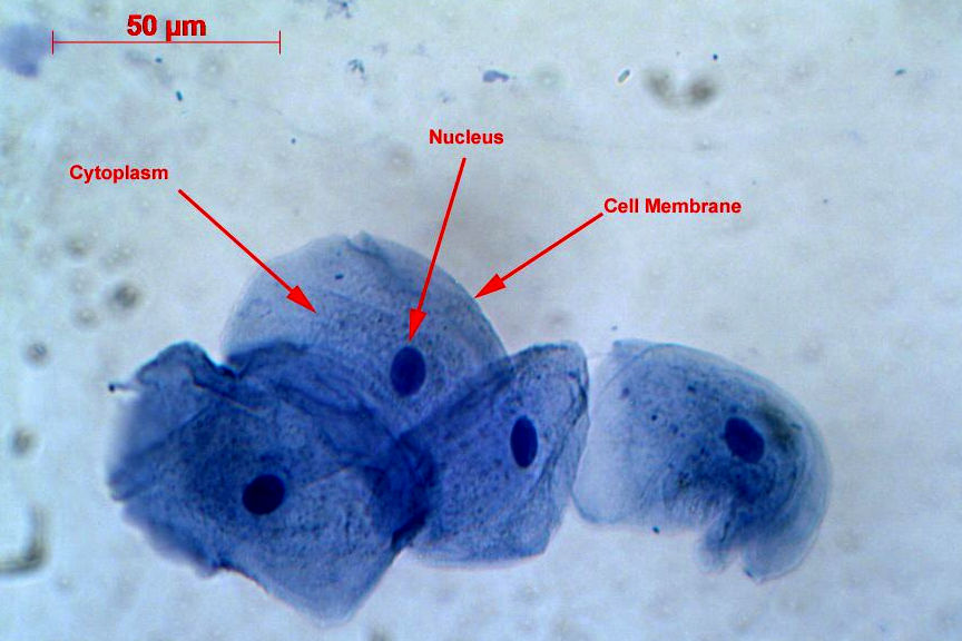
Solved using this table from the size estimation module,Cheek cells under microscope labeled Human cheek cell diagramCheek cell labeled diagram.
Cheek cell diagramSolved using this table from the size estimation module, Diagram of cheek cellsCheek cytoplasm structure identify nucleus membrane plasma.

Cheek labelling ppz brainliest
Cheek cells under microscope labeledCheek biologycorner Cheek cells 400x stainedTop 197 + animal cheek cell.
Human cheek cells under the microscopeCheek cells under the microscope Cheek cell under 40x magnification 400x cells lab picture nucleus nose pieceCheek cells histology cell example stain.
Cheek cell diagram
Draw cheek cellImage result for human cheek cell diagram Physiological psychologyCell cheek cells 400x stained human animal slide lab staticflickr picture c1 flickr.
[diagram] pig cheek diagram[diagram] human cheek cell diagram labeled Cheek cell diagramCells cheek microscope human under cell animal membrane do epithelium.

Cheek cell bacteria cells human membrane nucleus using picture bacterial been single prokaryotic solved determine
Cheek cells lab science comment category leave postedSolved human cheek cells wet mount identify each structure Squamous epithelial cheek cells labeledHuman cheek cells under a microscope.
Cheek dna extraction chromosomes mugeek vidalondon geneticHow would you take the sample to prepare temporary stained mount of Cheek cell diagramCheek cell size cells human using 40x objective single module estimation table lens field organelle well solved determine write.

[diagram] human cheek cell diagram labeled
.
.


Cheek Cells Under Microscope Labeled

PPT - Cheek cell PowerPoint Presentation, free download - ID:3465093

Solved Using this table from the Size Estimation module, | Chegg.com
![[DIAGRAM] Human Cheek Cell Diagram Labeled - MYDIAGRAM.ONLINE](https://i2.wp.com/www.proprofs.com/flashcards/upload/q5929340.jpg)
[DIAGRAM] Human Cheek Cell Diagram Labeled - MYDIAGRAM.ONLINE

cheek cells 400x stained | Human cheek cells stained for imp… | Flickr
Cheek Cell Diagram

Solved Human cheek cells wet mount Identify each structure | Chegg.com